Saturday, November 9, 2013

A very high carbohydrate diet for the reduction of elevated non-HDL cholesterol

Abstract


Public health authorities have for many years recommended diets high in complex carbohydrates for weight loss and prevention of heart disease. However, the research literature does not uniformly support the view that a replacement of fats, including saturated fats, with carbohydrates in the diet necessarily results in beneficial changes in cholesterol levels or heart disease risk. While very low carbohydrate diets have sometimes been observed to result in favorable changes to cardiovascular risk factors (due to the increases in HDL and decreases in fasting triglycerides often observed on those diets), there have been reports that, in a subset of the population, a very low carbohydrate diet may result in large increases in potentially atherogenic non-HDL cholesterol.

The reported studies to date have not been designed to investigate what happens to an individual with high non-HDL cholesterol who transitions from a long-term very low carbohydrate diet to a very high carbohydrate, non-vegetarian diet. The present study was designed to address that question using the author as the sole subject.

Results: Transition from a very low carbohydrate diet to a very high carbohydrate diet resulted in a rapid and dramatic reduction in non-HDL cholesterol. Improvements were also seen in oxidized lipoproteins, uric acid, and postprandial fat and carbohydrate metabolism. Seasonal allergies, which were virtually eliminated on the very low carbohydrate diet, returned upon adoption of the very high carbohydrate diet. No other deleterious effects were observed other than an increase in homocysteine, which was reversed through B-vitamin supplementation, suggesting the diet as implemented provided inadequate B vitamins. The diet is inexpensive and sustainable, though long-term effects (beyond 7 months) are not yet known.


The short version


You can watch my talk about this experiment at the New York Quantified Self meetup on Stephen Dean's Vimeo page. Note that this talk was given before I received my follow-up blood work showing the normalization of my elevated homocysteine and inflammatory markers.

Introduction


The present study was designed to measure the effects, primarily on blood lipids, of a 4-month very high carbohydrate, non-vegetarian dietary intervention (>65% carbohydrates on average) following several years of consumption of a very low carbohydrate diet, under approximately isoenergetic conditions (i.e. the intervention was adjusted to preserve pre-intervention body weight).

The study measured HDL and non-HDL cholesterol and a variety of other biomarkers. Note that, while blood lipids may be considered "risk factors" for heart disease, changes in these numbers do not necessarily represent a change in actual risk for heart disease. This study was not designed to detect changes in actual heart disease (which I don't have), and therefore I will say no more about actual heart disease in this write-up.

Conventional wisdom on carbohydrate consumption


Mainstream health authorities typically recommend a high level of dietary complex carbohydrate consumption. For example, the DASH diet (Dietary Approaches to Stop Hypertension) has been reported as including approximately 58% calories from carbohydrates (Swain et al 2011 “Characteristics of the diet patterns tested in the optimal macronutrient intake trial to prevent heart disease (OmniHeart): options for a heart-healthy diet”) and the TLC (Therapeutic Lifestyle Changes) diet recommends between 50% and 60% carbohydrate (Doucette and Kren, “The efficacy of using the Therapeutic Lifestyle Changes diet for reducing comorbidities associated with overweight and obesity”).

Some scientific research has cast doubt on the benefits of high carbohydrate consumption. For example, Walter Willett of the Harvard School of Public Health has argued that the substitution of saturated fat for carbohydrates is neutral from the perspective of heart disease risk. Based on a variety of dietary intervention studies, Willett argues that a decrease in saturated fat and a corresponding increase in dietary carbohydrate should result in an increase in fasting triglycerides and a decrease in HDL cholesterol (see e.g. Baum et al, "Fatty acids in cardiovascular health and disease: A comprehensive update"). These changes, which are considered deleterious, should compensate from the perspective of heart disease risk for the increase in non-HDL cholesterol, if any, that may be associated with the consumption of saturated fat. Other research implicates carbohydrates as a causative factor in the development of small, dense LDL particles, which are argued to be especially atherogenic (see this writeup at ketotic.org for a summary of this research).

In addition to potentially deleterious changes in HDL and triglyceride levels, advocates of low carbohydrate diets argue that consumption of a high carbohydrate diet will result in dangerous spikes in blood sugar as large quantities of carbohydrates are broken down to glucose and absorbed into the bloodstream (see, e.g. Jimmy Moore, Cholesterol Clarity, page 214, quoting Dr. Dominic D'Agostino).

Finally, research by Sharman et al (which I summarized previously) suggests that a high carbohydrate diet could cause deleterious changes in postprandial fat metabolism.

Because of this pre-existing research, this study was also designed to test the effects of the dietary intervention on postprandial blood sugar and triglyceride levels.


Review of a few long-term dietary interventions


Let's say you are an astronomer. You are working on a project that requires a long term observation of a particular celestial object. So you program your telescope to collect a year's worth of data on the object only to discover, at the end of the year, that the telescope had been looking at the wrong part of the sky. So do you analyze and publish the data you have, or do you start over and make sure your telescope is looking at what you wanted to study in the first place?

Now imagine you are a diet researcher...

I reviewed a sampling of dietary intervention trials lasting 12 months or longer to see what, if anything, they say about very high carbohydrate diets versus very low carbohydrate diets. This was based on a quick search and should not be considered comprehensive review.

I did not find the reported research to be terribly useful for the present study. With the exception of a series of papers examining the very low fat Ornish diet, none of the studies seemed to achieve a large enough difference in macronutrient intake between the different groups study participants (or between the study participants at baseline and at the end of the intervention) for me to consider them relevant to my experiment (which involved a change in fat consumption from approximately 60% to approximately 10%, excluding fat from fish). (Note: I excluded a number of studies by Caldwell Esselstyn because of his aggressive use of cholesterol-lowering drugs).

The table below shows the percentages of fat consumption in highest vs. lowest fat consuming study subjects. In cases where there was no control group, the baseline diet is used for comparison. Diet-induced changes in HDL and LDL cholesterol are also noted. I did not summarize changes in triglycerides but they generally show the same trends as HDL – studies that showed an increase in HDL generally showed a decrease in fasting triglycerides.


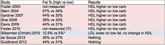
Summary of changes in HDL and non-HDL cholesterol at conclusion of selected long-term dietary intervention studies. *Silberman et al fat consumption percentage was calculated from reported grams of fat consumed per day assuming a 2,000 calorie diet.

References: Foster 2003Stern 2004Gardner 2007, Shai 2008Davis 2009Foster 2010Silberman 2010de Souza 2012Guldrand 2012.

Note: the Silberman (Ornish) subjects started out on a very low fat diet, and they transitioned to a diet much lower in fat. Even their starting level of fat consumption is far lower than anything achieved in the other “low fat” interventions summarized above.

The other studies could charitably be described as at best “mildly effective” in achieving their dietary objectives. The numbers shown in the table above for final macronutrient ratios are generally based on surveys conducted on the participants at the conclusion of the study (except for Foster, who did not survey the dieters in either study and therefore apparently doesn't know what the subjects were actually eating). There is a rather telling comment in de Souza et al 2012"despite the intensive behavioral counseling in our study, macronutrient targets were not fully met, which complicated the interpretation of our null result." So they told different groups of people to eat different diets, but they all ate basically the same diet. Their outcome measures did not differ between groups at the end of the study (the “null result”), and therefore interpreting the data is “complicated.” Let me suggest, actually, that interpreting their data is "a waste of time." (They published it anyway, of course.)

By the way, the Silberman study on the Ornish diet had 2,974 people in the intervention group (it was not a controlled trial). It is interesting that the Ornish researchers appear to be able to get people to actually eat very low fat diets, while other researchers seem to have more trouble getting participants to make such dramatic diet and lifestyle changes. I'm not commenting one way or the other on the Ornish plan, but it is a bit disappointing that the other research groups don't seem to be able to effect such large changes in macronutrient intake in their study participants. This means the published studies are not especially helpful in evaluating diets at the extreme ends of the macronutrient spectrum.

A number of the Ornish studies observed short term reductions in HDL. However, the longer studies seem to indicate that those HDLs rise again over the long term (3-5 year timeframe). What is potentially more troubling, however, is that the Ornish studies do not seem to report a meaningful reduction in fasting triglycerides.

In 2004, Yancy et al ran a study of a very low carbohydrate ketogenic diet for 24 weeks. Two of the subjects (out of 59) on the low carbohydrate diet dropped out because of sudden increases in non-HDL cholesterol. Overall, 30% of the subjects on the very low carbohydrate diet experienced an increase in LDL cholesterol of 10% or more, compared to 16% of subjects on the low fat diet (this difference was not statistically significant). Because of its short duration, this study did not qualify for inclusion in the summaries above. However, it does support the hypothesis that a very low carbohydrate diet can raise LDL in a minority of the people who try it (unfortunately Yancy et al did not report non-HDL levels in these individuals, which would have been much more useful). This is also supported by anecdotal reports from individuals consuming very low carbohydrate diets. As far as I know a study designed to test this hypothesis has not been conducted.


Fish oil studies


A number of studies have investigated the effects of fish oil supplementation on risk of cardiovascular disease. These have not always found fish oil to be beneficial (see e.g. Risk and Prevention Study Collaborative Group "n-3 fatty acids in patients with multiple cardiovascular risk factors" finding no benefit for cardiovascular mortality or morbidity). However, these studies generally involve very low doses of fish oils, on the order of 1 gram of total n-3 fatty acids per day. A study will find no benefit if it uses an intervention that is too small, but this of course tells you nothing about the effects of a larger dose.

Some studies using a larger dose (e.g. Harris et. al. Journal of Lipid Research 1988, which used 24-28g omega-3 per day, and Phillipson et. al., New England Journal of Medicine 1985, which used approximately 20-25g omega-3 per day) have shown a dramatic improvement in metabolic markers, including total and non-HDL cholesterol, but these studies were short term and not designed to observe changes in heart disease. Based on this I believe it is more likely than not that a dose sufficient to improve metabolic markers is likely to also have beneficial effects against heart disease. The present intervention involves a very large intake of n-3 fatty acids from fish.


Dietary cholesterol recommendations


It is commonly heard that dietary cholesterol has at most a small relationship to blood cholesterol levels. This seems to be the case when cholesterol intake is high at baseline. For example, Ancel Keys suggested that a reduction in dietary cholesterol from 600 mg to 300 mg per day on a 2,000 cal/day diet would be expected to result in a reduction in total serum cholesterol of only 7.6 mg/dl ("Serumcholesterol response to dietary cholesterol," American Journal of Clinical Nutrition 1984). According to Keys, the relationship between dietary cholesterol and serum cholesterol is stronger at lower levels of dietary cholesterol intake. Regardless of the strength of this relationship, public health authorities continue to recommend a reduced cholesterol diet as a preventive measure for cardiovascular disease. The recommendation in the 2010 DietaryGuidelines for Americans is <300mg/day.

The figure below is reproduced from Endocrinology and Metabolism, Third Edition (Felig, Baxter and Frohman, McGraw Hill 1995, page 1368). It shows (hypothetically, I presume) the relationship between dietary cholesterol and serum cholesterol. Consistent with the Ancel Keys paper cited above, the curve has a decreasing slope as dietary cholesterol increases, eventually leveling out. This sort of pattern might be expected with a regulated biological process, where the body seeks to maintain serum cholesterol at a particular level regardless of input. In that case, the "ceiling," where the curve flattens out, may tell us something about what the regulatory system is trying to achieve.


Personal motivation


Why did I do this? I have been tracking my cholesterol levels over the past few years since they have been generally higher than what is considered normal by mainstream medical opinion (without making any judgements about the validity of that opinion). In addition, since adopting a low carbohydrate diet in 2009, I have observed a slow but persistent trend towards increased total and non-HDL cholesterol. Therefore, I have tried a number of interventions to bring those numbers down. My intention is not to allow the blood lipid numbers to dictate my dietary choices. However, I believe an understanding how diet affects my blood lipids is useful information for making better choices about what to eat. I'd like to take into account all potentially relevant information.

I first tried a low carbohydrate diet after reading Good Calories, BadCalories, just to see what would happen. It caused a number of health improvements right away. During this time I noticed (with a glucometer) blood sugar spikes after carbohydrate-containing meals and was not sure if they were within a healthy range. I stayed on the low carb diet because I felt fine and it seemed to have improved my health. However, I had never tried a very high-carbohydrate diet and wanted to see what would happen.


Hypotheses


This study was designed to test the following hypotheses:
  1. A high carbohydrate, low fat diet can meaningfully reduce non-HDL cholesterol
  2. An increase in dietary carbohydrate lowers HDL and raises fasting triglycerides
  3. High carbohydrate diets cause excessive spikes in blood glucose throughout the day
  4. High carbohydrate diets impair postprandial triglycerides after an oral fat tolerance test
The diet, timeline and measurement protocol were designed to evaluate these hypotheses. Based on prior review of the scientific literature, I thought the first hypothesis was false and the others were true.


Design and Methods


The study consisted of a single dietary intervention phase conducted after long-term consumption of a very low carbohydrate baseline diet (total carbohydrate intake averaging less than 75g/day).

Approval of an Institutional Review Board was not required for this n=1 self-experiment. The author's mother and girlfriend were informed of the study design in passing and they raised no ethical concerns. The study was conducted according to ad hoc human subjects research guidelines made up on the spot by the author, and reviewed and approved by the author as the sole human subject.


Baseline diet


The baseline diet consisted primarily of whole eggs (3-4/day), grass-fed red meat (450g/day on average), butter (1/2 stick/day on average), almonds (30g/day on average), non-starchy vegetables, and coffee (2 cups/day). For approximately four weeks before the start of the intervention phase, carbohydrate consumption was gradually increased to approximately 150g/day by the addition of bananas to the baseline diet.


The intervention diet


The intervention diet consisted primarily of white basmati rice (Swad "premium quality" Dehraduni aged basmati rice) and frozen wild coho or sockeye salmon (Trader Joe's). In addition, a typical day included approximately one bunch of bananas (1-2 pounds), 9.5 oz of grass fed whole milk yoghurt (Grazin' AngusFarms), 1 oz almonds, some sort of shellfish once or twice a week, and a variety of green vegetables. A few meals a week would be at restaurants and consist of whatever I wanted. The amount of rice consumed varied to meet caloric needs and varied between approximately 450g and 565g (dry). Target vegetable intake was determined to roughly meet vitamin requirements according to US daily reference intakes, but in practice the requisite amount of green vegetables was often not achieved. During peach, apricot, and cantaloupe season here in the Northeast U.S., I ate, respectively, a lot of peaches, apricots and cantaloups.


1.75 pounds of white basmati rice. 1 pound of fish.

Carbohydrates: I was looking for a relatively low-glycemic carbohydrate source. I thought I would avoid sweet potatoes as they had appeared to lower myHDL in a prior short-term experiment. So I went with white rice, a common global staple food. Basmati rice is reputed to have a low glycemic index relative to other forms of rice, and I live a few blocks from a South Asian neighborhood and therefore have a convenient supply of high quality Indian rice in ten pound bags.

According to my Endocrinology and Metabolism textbook (Felix, Baxter and Frohman, 3rd Ed.), the increase in fasting triglycerides and corresponding decrease in HDL commonly observed in a high carbohydrate dietary intervention occurs only when carbohydrate intake is increased abruptly, and does not occur with a gradual transition period (see page 1372). Therefore, the present study utilized a wash-in period of several weeks during which carbohydrate consumption was increased gradually from ~75g/day to ~150g/day.

Protein: The intervention diet was designed to have approximately the same protein content as the baseline very low carbohydrate diet. I use the “one gram of protein per pound of body weight” rule of thumb which is widely followed for active individuals looking to build or maintain muscle mass (approximately 150g/day). Given the somewhat mixed evidence on dietary cholesterol, I wanted to try keeping cholesterol intake relatively low while obtaining this amount of protein. Therefore, fish (primarily salmon and trout) was chosen as a compromise between cholesterol content and high-quality, whole food protein. Because of the target protein consumption, cholesterol intake somewhat exceeded the mainstream guidelines for cholesterol of 300mg per day (see the 2010 Dietary Guidelines for Americans). Since I was aiming to achieve my target protein requirements by eating fish, I did not need to eat any of the "protein" sources such as tofu, quinoa, beans, etc. that are commonly consumed on other low fat and vegetarian diets.

Fiber: The diet as implemented is relatively low in fiber. I briefly looked into the research on fiber and did not feel compelled to go out of my way to consume it. Because of that I chose white rice as my staple carbohydrate instead of brown.


Measurements


Periodic measurements of total and HDL cholesterol were taken with a CardioChek PA meter. In addition, after 8 weeks of the intervention diet, a comprehensive blood and urine analysis was performed, including Atherotech VAP lipoprotein testing (Shiel Medical Laboratories, Brooklyn, NY) and compared with a similar panel taken one year prior during the baseline diet (high in red meat, butter and green vegetables but excluding grains, legumes and non-butter dairy).

Postprandial testing


After adaptation to the very high carbohydrate diet for at least 8 weeks, I conducted a number of postprandial tests. First was a standard oral glucose tolerance test using 75 grams of glucose (Kalustyan's, New York, NY) dissolved in New York City tap water.

I also attempted a “real food” torture test by adding a 9" cantaloupe to a typical dinner of wild salmon. I have no idea how much glucose was in that particular cantaloupe but I believe it must have been substantially more than 75 grams. In order to simulate “worst case” conditions, I wolfed it down as fast as possible, which took about 10 minutes.

In order to test my hypothesis about the effects of a very high carbohydrate diet on postprandial triglycerides, I conducted an oral fat tolerance test based on a typical breakfast I consumed during the last year of my low carbohydrate diet. This consisted of four eggs (Grazin' Angus Farms) cooked (over easy) in coconut oil, plus half a stick of butter. This is more fat, more saturated fat and more cholesterol than is typically used for oral fat tolerance tests in research settings, though contrary to most researchers I did not include any carbohydrates (or wheat) in my test. For these reasons my results will not be directly comparable to any oral fat tolerance test from the research literature (which is just as well, because, due to lack of standardization, published results are rarely comparable to each other). However it does have the virtue of being directly comparable to oral fat tolerance tests I have performed on myself and written about before. I have noticed previously that triglycerides after a meal may be very low on the day after heavy exercise. Therefore I conducted my oral fat tolerance test for this experiment on a day after a day on which no heavy exercise was performed.


Analysis


Results were recorded using the iPhone Notes app and bits of paper and plotted in R. Statistical analysis was not considered necessary or useful for this experiment. I also did not need WiFi, Bluetooth, a proprietary machine learning algorithm, The Cloud, Web 2.0, or any other fancy technology.


Results


Executive Summary


I observed the following changes on the intervention diet compared to baseline:
  1. Very large decrease in non-HDL cholesterol, LDL cholesterol and oxidized lipoproteins
  2. No change in HDL cholesterol or fasting triglycerides
  3. decrease in serum uric acid
  4. No adverse postprandial responses to high carbohydrate or high fat meals
  5. Seasonal allergies returned
  6. Intervention diet (as implemented) may be insufficient in B vitamins


Cholesterol levels


The figures below show my non-HDL and HDL cholesterol levels during the baseline (low carbohydrate, red) and intervention (high carbohydrate, blue) diets. The reduction in non-HDL was immediately evident by the first measurement, which was taken after only 7 days on the high carbohydrate diet. No clinically meaningful change is evident in HDL cholesterol.


Non-HDL cholesterol on baseline (low carbohydrate, red) and intervention (high carbohydrate, blue) diets.

HDL cholesterol on baseline (low carbohydrate, red) and intervention (high carbohydrate, blue) diets. The increase in the 2.5-3.5 year period roughly corresponds with high butter consumption. Note the downtrend towards the later part of the high-butter period.


Fasting triglycerides were essentially unchanged (57 on 4/3/2012 to 63 on 5/31/2013).


Advanced lipid testing


Direct LDL measurements performed on April 3, 2012 (on the baseline diet) and again on May 31, 2013 (after 8 weeks on the very high carbohydrate diet) revealed a decrease in total LDL (direct measurement via the Atherotech VAP) from 190 mg/dl to 77 mg/dl.

Results of advanced cholesterol testing (Atherotech VAP).


Oxidized LDL and HDL


Along with the decrease in non-HDL cholesterol, oxidized LDL decreased from 62 to 35 mg/dl and oxidized HDL decreased from 36 to 19.


Blood sugar control


The figure below shows the results of an oral glucose tolerance test done on the morning of June 7, 2013. My blood sugar reached a peak of 152 at 45 minutes and returned to baseline within 2 hours.

Blood sugar in response to an oral glucose tolerance test containing 75 grams of Kalustyan's glucose dissolved in New York City tap water.

The figure below shows my blood sugar over most of a typical day (in this case, May 28, 2013). The majority of my carbohydrate consumption was in the late morning and over lunch (12-1 pm). For reference, approximately 4 bananas and two pounds (cooked) of basmati rice were consumed before 1 pm. As you can see, no abnormally high or low blood sugar levels were observed. The highest reading for the day was 126 mg/dl.


Blood sugar readings over the course of a typical day on a very high carbohydrate diet.


Hemoglobin a1c is a measure of glycated hemoglobin. It varies from person to person and may also depend on average lifespan of red blood cells, so it has some limitations as a biomarker, but it is considered a useful measure of heart disease risk, to the extent that it may be mediated by long-term elevations in blood sugar. This year, after two months on the high carbohydrate diet, my hemoglobin a1c was ever so slightly lower than it has been previously on the low carbohydrate diet (5.6% on 4/3/2012 vs. 5.5% on 5/31/2013).

The standardized 9” oral cantaloupe tolerance test resulted in a maximum postprandial blood sugar of 107.


Triglycerides


Below are the results of an oral fat tolerance test conducted on July 30, 2013 according to the protocol described above.

Triglycerides before (t=0) and after (t=150 and 210 minutes) a high fat test meal consisting of four eggs, five tablespoons of coconut oil and 1/2 a stick of butter. The peak value of 111 mg/dl occurred at 150 minutes.


Allergies and hives


One of the clearest benefits I noticed when I started eating a very low carbohydrate diet was a sharp reduction in my seasonal allergies. On the very high carbohydrate intervention diet, my spring allergies returned. In addition, over the first 3 weeks of the diet, I started getting hives. The hives went away after the first three weeks, and so have the allergies. The allergies returned during the fall allergy season (October).


Uric acid


One unexpected benefit of the very high carbohydrate diet was a reduction in serum uric acid, from a slightly high 8.3 mg/dl on 4/3/2012 to 6.8 on 5/31/2013. I have not investigated the likely cause or meaning of this change, but my lab defines the reference range as 4.0-8.0 mg/dl, and elevated uric acid levels are associated with impaired kidney function.


Homocysteine and c-reactive protein


Initially, an increase in homocysteine and c-reactive protein was observed (as of 5/31/2013). Elevation in homocysteine may have been related to a deficiency in B vitamins and supplementation was commenced (25mg B6, 2,000 mcg B12 and 1,600 mcg methyl-folate). Elevation in c-reacitve protein is believed to be caused by a minor viral infection at the time the May 2013 blood work was conducted.

Homocysteine and c-reactive protein were retested and confirmed within normal range on 8/23/2013.


Discussion


Interventions that make small changes to macronutrient composition may be expected to result in small changes in blood lipids. Studies like that require statistical analysis with n>>1 to reasonably reject the null hypothesis that a particular dietary intervention results in no change, or no improvement, in health or biomarkers. The present study was designed to induce a large change in blood lipids by way of a very large change in macronutrient intake. As with all diet studies, it necessarily involved a change in multiple dietary factors as certain foods were reduced or eliminated (e.g. red meat), and others were increased (e.g. fish). Therefore, it is not possible to determine whether the effects observed were the result of changes in macronutrient content, or of other concurrent changes.

It seems reasonable to assume that the effects of macronutrient changes, if any, may not be linear. For example, it may not be possible to infer the effects of a diet comprised of 65% carbohydrates from a study population consuming no more than 55% carbohydrates on average. This fact may help explain the results of the dietary intervention studies, where the only interventions involving fat consumption below 10% of calories (the Ornish studies) were able to demonstrate decreases in non-HDL cholesterol. In addition, studies are usually not designed to detect instances where a subset of the population shows an unusually large response to one intervention or another.

Contrary to my initial assumptions, this experiment strongly supported the hypothesis that a very high carbohydrate diet can lower non-HDL cholesterol. In addition, it failed to support the hypotheses that high carbohydrate diets lower HDL, raise triglycerides, cause unhealthy blood glucose spikes and impair oral fat tolerance. Again, it may be the case that these effects occur only in a subset of the population, but this hypothesis has not been confirmed or refuted because of the design of the dietary intervention studies I reviewed.


Fasting measurements


My HDL levels on the very high carbohydrate diet were consistent with their levels during the first few years of the low carbohydrate diet, prior to the year of high butter consumption. However, given the study design (n=1) and the natural variability in cholesterol levels from day to day, this study is not powered to detect small decreases in HDL. And why would I want to detect a very small decrease in HDL? A small decrease most likely won't make any difference to me personally. I had previously conducted a 4-week study of the effects of adding sweet potatoes to a very low carbohydrate diet. I observed a decrease in HDL during this time which was reversed once the sweet potatoes were removed. My current results are not consistent with that finding, or with other results (unpublished) suggesting that my postprandial triglycerides are adversely affected by carbohydrate consumption.

A number of plausible solutions to this conflict are i) certain carbohydrates (e.g. sweet potatoes  adversely affect HDL and triglycerides, while others (e.g. white rice) do not; ii) carbohydrates lower HDL and raise fasting triglycerides when eaten with fat, but not in the context of a very high carbohydrate diet where fat intake is low; iii) high fish consumption counteracts any adverse effect on HDL and triglycerides that would otherwise have occurred; and/or iv) as suggested by my Endocrinology and Metabolism textbook, the several week wash-in period during which carbohydrate consumption was gradually increased was effective in preventing these adverse changes.

High carbohydrate diets are often claimed to cause deleterious changes in LDL particle size. However, in my case, advanced lipid testing performed on May 31, 2013 reveals favorable changes in all lipoprotein subtractions. Total small, dense LDL particles (LDL 3 and LDL 4 on the VAP test) decreased from 99 mg/dl on April 3, 2012 (on the baseline low carbohydrate diet) to 37 on May 31, 2013 (8 weeks into the very high carbohydrate diet). Larger LDL subtractions also decreased but by a smaller absolute and relative amount (91 to 40). Therefore, the dietary intervention has apparently caused a favorable shift in both the ratio of large vs. small LDL particles, and also in the absolute amount of small, dense LDL. There was also a small decrease in VLDL, from 16 to 14 mg/dl.

There was also a slight favorable shift in HDL subfractions. While the total HDL cholesterol was essentially unchanged (68 mg/dl on 4/3/2012 to 69 on 5/31/2013), the balance between large/buoyant HDL 2 (believed to be most protective) and the small/dense HDL-3 shifted from 19/49 to 22/46. However, this change is small and it is not clear if it has any clinical relevance.

My measurements of oxidized lipoproteins also contradict a common belief in low-carbohydrate diet communities: that reduction in carbohydrate consumption will reduce lipoprotein oxidation and therefore reduce heart disease risk regardless in changes in total lipid levels. (See, for example, the quote from Jefrey Gerber on page 87 of Jimmy Moore's Cholesterol Clarity). He says that lowering carbohydrates will lower oxidation, but my oxidized lipids decreased enormously on this diet. In addition, while the decrease in oxidized LDL would be expected given the large decrease in total LDL, there was also a large decrease in oxidized HDL despite total HDL levels remaining essentially unchanged. While the absolute level of oxidized LDL decreased from 62 to 35, on a relative basis as a percentage of total (direct) LDL, it increased from 33% to 45%. Oxidized HDL decreased on a percentage basis from 53% to 28%.

The elevation in homocysteine suggests that the diet as implemented provided inadequate B vitamins. Although the design of the diet included a substantial amount of B vitamin-containing green vegetables, the diet as implemented did not. Supplementation (25 mg B6, 1600 mcg methyl-folate and 2000 mcg B12) rapidly reversed the adverse change in homocysteine.


Postprandial measurements


Because of the human body's ability to adapt to a wide variety of diets, I had assumed at the outset that improvements in postprandial blood sugar control may occur in response to the very high carbohydrate diet, and that this would likely produce a normal oral glucose tolerance test response. In fact my glucose tolerance test results on the very high carbohydrate diet are considered to be within normal standards. Note that, although the 2-hour reading (70) is lower than the fasting level, I was not at any time symptomatic of hypoglycemia.

You can contrast the oral glucose tolerance test result with the full day blood sugar measurements. Even though the glucose tolerance test involved the consumption of a much lower dose of carbohydrates (75g), it produced a dramatically higher blood sugar excursion than the worst case seen during a full day (500g carbohydrates). This suggests that the oral glucose tolerance test is not representative of an actual day of very high carbohydrate eating (though perhaps it may be representative of junk food or soft drink consumption).

Some people are afraid to eat fruit these days because of concerns about blood sugar. My postprandial blood sugar after the oral cantaloupe tolerance test peaked at 107, so I'll say with confidence that I am not likely to run my blood sugar up to unhealthy levels while eating real foods. Note that the protein consumed along with the cantaloupe likely triggered an insulin response that could have reduced the peak blood sugar level.

Left: the aftermath of a standardized 9” oral cantaloupe tolerance test. My peak blood sugar of 107 is shown on the glucometer. The cantaloupe was very ripe and delicious.

Right: 75 grams of glucose. Yikes!


Eating a very high carbohydrate diet might be expected to lower your postprandial response to carbohydrates. However, it might also be expected to worsen your postprandial response to fats, because a very high carbohydrate diet is necessarily very low in fat.

On the very low carbohydrate diet, my peak triglycerides after a typical breakfast (described above) would occur around 3.5 hours after the meal and would usually reach approximately 155 mg/dl. On some days, particularly if I had done some extremely heavy exercise the day before, my peak triglycerides would reach only 100 mg/dl.

On the very high carbohydrate diet, my triglycerides after this test meal stayed admirably low (111 mg/dl). Although it is impossible to draw firm conclusions from a single test (since peak postprandial triglyceride levels can vary significantly from day to day and the reasons for this variability are not entirely clear), this is nevertheless a surprising result. Based on my prior research, I was expecting my oral fat tolerance to be impaired on the very high carbohydrate diet, and that this would be evidenced by a higher and possible also a later peak reading. If anything, this result suggests an improvement in oral fat tolerance. The results, taken together, therefore suggests a true improvement in metabolism with no observable metabolic downsides.


Odds and ends


The return of my allergies on the very high carbohydrate diet was not entirely unexpected, because I had suffered from seasonal allergies fer years prior to adopting the low carbohydrate diet. They ended after a few weeks, which may have been due to the end of allergy season, or possibly because of a quercetin/bromelain supplement suggested by my doctor. My typical October seasonal allergies also returned, and also may have responded to the same supplement. At this point it is impossible for me to separate the effects of the supplement from the end of each allergy season. The hives were unexpected, but temporary and I have no reason to think they will come back.

One of my concerns in transitioning to a very high carbohydrate diet was with my teeth. However, I have not noticed any increase in root sensitivity or other adverse dental health effects.


Competing financial interest disclosure


The author does not declare any competing financial interests. The author also declares affirmatively that he has no competing financial interests related to this research that an ethical person would feel ethically obligated to declare.


Conclusions


The present study demonstrated a dramatic reduction in non-HDL cholesterol in a short period of time in connection with the adoption of a very high carbohydrate, non-vegetarian diet. Improvements were also seen in oxidized lipoproteins, uric acid, and postprandial fat and carbohydrate metabolism. Seasonal allergies, which were virtually eliminated on the very low carbohydrate diet, returned upon adoption of the very high carbohydrate diet. No other deleterious effects were observed other than an increase in homocysteine which was reversed through B-vitamin supplementation, suggesting the diet as implemented provides inadequate B vitamins. The diet is inexpensive and sustainable, though long-term effects (post 7-months) are not yet known.

Saturday, July 20, 2013

Apricot-braised Trout

In honor of apricot season, here is something you might want to do with a bunch of extra apricots -- braise fish in them! Trout is not the mildest of fish, but the other flavors in this dish, particularly the cloves and braised apricots, combine to impart an almost meaty quality. The spices add body and complexity to the sauce. A lesser fish might be lost in the fray.

I had leftover fresh baby fennel from the weekend that I wanted to use, but I'm on the fence about it here and I think the dish might work just fine without it. Don't get me wrong -- it was very good, but I wouldn't have fresh baby fennel air-dropped from Lebanon just so you can make this.

The fish are braised whole. However, you should consider removing the bones and spine while they are raw, as the braised meat is very soft and more difficult to separate from the bones than a roasted fish would be. Please save the bones (and heads) to make stock (or send them to me -- I have an idea for an improved fish popsicle).

For cookware all you will need is a skillet with a reasonably well-fitting cover for your preferred braising method. I like to braise on an induction cooker, which won't heat up your kitchen in the summer as much as an oven or gas burner.

Kneeless Apricot-braised Brook Trout


Ingredients:
  • two brook trout (approximately 1 pound each), whole, cleaned and scaled
  • flesh from four or five apricots, cut into wedges
  • one stalk of fresh baby fennel (use the white portion, compost the greens)
  • onions
  • 1-2 tsp salt and ground pepper to taste
  • 5 whole cloves
  • 2 pats of butter (1/4 stick)
Spice mix:
  • 1/2 tsp ground cumin
  • 1/2 tsp ground coriander
  • pinch of ground cayenne pepper
Steps:
  1. in a skillet, sauté onion and cloves in butter until the onions brown slightly
  2. add fennel and sauté until soft
  3. add spice mix and continue to sauté. Spices will darken slightly and start to form a paste
  4. add apricots and trout, salt to taste, and grind black pepper on top
  5. add 1/4 to 1/2 cup of water (some more will come out of the apricots), cover, and braise on low heat until fish is cooked through (30 minutes should do it but don't worry if you go a bit over)
Kneeless Apricot-braised Trout

I consulted professional wine consultant Shana Reade and she provided the following suggestions.

My first inclination would be to suggest a Alsatian Gewürztraminer. They are typically fruity, floral, and a bit spicy. I always taste clove in the good ones, so that would pick up that note in the fish nicely. It's also a bit of a heftier wine, so wouldn't be overwhelmed by the richness of the trout. Sidebar: this wine might be the flavor you are looking for in the cod pops instead of using dextrose. I will see if I have a bottle laying around the office. 
Some people are put off by any sort of residual sugar (when fermentation is stopped prior to the yeast consuming all the sugar in the grapes, so there will be more sweetness) in a wine, and Alsatian Gewürz's might have a touch in addition to already having lush tropical fruit flavors, so the dry wine fans might like a Chenin Blanc from the Loire. The two main appellations are Savennieres or Vouvray. Both would work, but Vouvray is made with varied levels of residual sugar, and they are really bad about telling people that on the label, so I think a Savennieres is the way to go, and it's awesome. It's usually a bit funky and yeasty, but mineral driven with touches of honey, and sometimes tastes a bit like wool or lanolin, but in a good way?

Now when you said esoteric, this wine came to mind (Movia Rebula). I don't think the description gives justice to how neat this wine is. It's almost an orange tinge, and seems racy and rich at the same time, and kind of like drinking a bouquet of dried flowers.
And even though this is probably going on far longer than you anticipated, for the red fans, I think a Beaujolais cru would be perfect. Beaujolais got a bad reputation for their Beaujolais nouveau, which uses a type of fermentation that produces a wine that tastes like bubble gum and banana bread. But there are 10 little villages, or crus, that make a lighter style red, with a firm but not aggressive structure, typically with a nice balance of fruit and earthiness.

Monday, April 15, 2013

How I fell down a murine rabbit hole

This post is about animal models and postprandial elephants.


Personal update


Before I get started on today's topic, I wanted to introduce my latest experiment. Although butter had a temporary beneficial effect on my cholesterol levels, the benefit did not persist. In fact, towards the end of last year, while my HDL levels remained quite high, the non-HDL crept up into territory I was not comfortable with. I can't say if this was due to the butter, a year of relatively high egg consumption (3/day), a longer term effect of high red meat consumption, or the consequence of several years on a low carbohydrate diet. Whatever the cause, I decided, since I've never done it before, to try out an actual high carbohydrate diet that is low in fat and cholesterol. At least at the end of this experiment I'll be able to speak about high carbohydrate diets from a position of personal experience.

I ramped up my carbohydrate consumption gradually over a couple of weeks, and then abruptly switched out all the butter, eggs and red meat for about a pound each of white rice, bananas, fish, and the occasional mound of white potatoes. I sprinkle in a sprinkling of almonds, salad greens, carrots, spinach, shellfish, yogurt, and whatever else I feel like eating to round out my micronutrient requirements, which I roughly confirm from the USDA data. Wheat is still out, as are the vegetable oils and all processed foods.

It is too early to report much in the way of results, but in the first two weeks my HDL has dropped a little, my fasting triglycerides stayed under 70, and my non-HDL has dropped about 120 points. What is also a bit of a surprise is that my blood sugar has been quite consistent and low, almost always peaking below 125 even following heroic quantities of white rice. One benefit of rice in regard to blood sugar is that I find it difficult to eat it quickly (compared to, say, a banana or sugar water). Anyway the new diet seems to agree with me so far at this very preliminary stage. In a few weeks I will feel more qualified to comment on high carbohydrate diets, having actually tried it for the first time in my life.

"Totally Misleading"


Now on to the meat of this post. Episode 52 of "This Week in Microbiology" featured a conversation with clinical microbiologist Ellen Jo Baron. Although it is not the focus of the episode, she briefly told the story of how difficult it was to publish her PHD thesis on salmonella. She says:

"What we did was compared in vitro neutrophil activity of mouse neutrophils against the mouse typhoid organism salmonella typhimurium, and then human neutrophil activity against the human typhoid agent salmonella typhi, and my work showed that they were radically different early responses, and nobody wanted to buy that because many of the premier researchers in the field had been using the mouse model of typhoid looking at vaccines et. cetera and I was showing them that it was an inappropriate model."

In other words, the mouse immune system behaved differently from the human immune system in what was supposed to be an animal model of a human condition. Established scientists did not like the finding and resisted its publication. However, This took place in the depths of time (the 1970s), so perhaps we can forgive our primitive forbears for their unfounded prejudices.

Or maybe not? Recently in the New York Times, Gina Kolata describes a similar episode in the life of a paper published by Seok et. al. this February in the Proceedings of the National Academy of Sciences.

Like Baron, Seok et. al. were studying the behavior of immune cells under an assortment of inflammatory conditions (namely sepsis, burns and blunt trauma). The new paper recorded patterns of gene expression in human white blood cells taken from real patients. According to the the Times, the researchers ran into trouble getting their results published because they had not demonstrated that their results were consistent with the established animal models for these conditions. So they ran the experiments again with mouse cells, and, lo, they did not match at all. This, apparently, did not comfort the paper's critics. Only by submitting to PNAS (along with a recommendation that specific reviewers be consulted) was the team able to win publication.

So back to the results: after an infection, burn, or getting traumatized by a blunt instrument, a wide range of changes occur over time in gene expression in white blood cells, as they participate in the immune/healing response. Expression of some genes will increase while expression of others will decrease. As between human cells and the corresponding mouse model, correlations (r-squared) max out at 8% in the directionality of gene expression. In other words, if a gene was upregulated in humans, it was essentially random whether it would be unregulated or downregulated in the corresponding mouse model. Just about half the time, the same gene, coding the same protein, under what is supposed to be the same stimulus, does the opposite thing.

Interestingly, while gene expression patterns in the three human conditions were quite similar to one another, the three mouse models were very different not only from the human conditions but also from each other. So the human inflammatory response is a maze of twisty passages, all alike, while the mouse inflammatory response is a maze of twisty passages, all different (and all different from the human twisty passages). In addition to the directional differences in gene expression, the researchers also observed enormous differences in timing. For example, a response that lasted 4 days in the mouse might persist for 6 months in people. Yikes!

These results led Gina Kolata of the Times to say that the mouse models for these conditions were "totally misleading." Seok et al, introduce their paper by pointing out that "there have been nearly 150 clinical trials testing candidate agents intended to block the inflammatory response in critically ill patients, and every one of these trials failed" (Seok page 1). (these words were too strong even for the Times -- they changed "every one" to "most"). This paper is very clearly written and I can't really sum it up better than they do:

"here, we show that, although acute inflammatory stresses from different etiologies result in highly similar genomic responses in humans, the responses in corresponding mouse models correlate poorly with the human conditions and also, one another. Among genes changed significantly in humans, the murine orthologs are close to random in matching their human counterparts."

So another strike against the mouse as being the perfect biochemical equivalent to a human.

Meanwhile it has been known for many years that mice respond very differently than humans to bacterial lipopolysaccharide (LPS). Also known as endotoxin, LPS has made a number of prior appearances on this blog. The hapless human is about 100,000 times more sensitive to LPS than the mouse. In 2010, Robert Munford published "Murine response to endotoxin: another dirty little secret?", citing work by H. Shaw Warren, the second author on the Seok et. al. paper. So the new gene expression results add to an existing body of evidence casting doubt on the presumed similarities of human and murine immune systems.

I guess, after all, rodents and humans are not so much alike. For reference, here's a recent mammalian family tree showing the distance between humans and rodents. (McCormack et al Genome Research 2012).

J. Craig Venter gets to appear personally on phylogenetic trees like this one.

The differences in gene expression between animals and humans in response to lipopolysaccharide and inflammation in general, from the 10^6 difference in sensitivity, time course differences of several orders of magnitude, and essentially zero correlation even in directionality, cast significant doubt on my earlier thoughts about lipopolysaccharide and postprandial triglycerides. As I read studies on this topic, I am constantly finding myself disappointed to pull up references only to find animal research. This is not always disclosed in the title of the paper, or even in the abstract! In my darkest hours I sometimes find myself pointing this out to people on twitter (by the way, if I can fit "in mice" in a 140 character tweet, you can fit it in the title of your peer-reviewed research paper).

Digression on (Mathematical) Models


Recently I picked up a very nice little text book by Hugo van den Berg on mathematical modeling of biological systems.  It covers some of the basic stuff that I think someone doing mathematical modeling should be expected to know. Since I have been spending more time reading a book and less time on the Internet, Google has backed down on sending me ads for modeling agencies. And all of the modeling agencies that twitter-followed me have since un-followed me.

Some readers may not know that the field of computational biology was founded by Alan Turing, the father of computer science himself. His landmark 1953 paper on morphogenesis is available online.


xkcd #793.


If you precisely define the characteristics of an imaginary object, and then investigate the consequences of your definition, you are doing mathematics. If your imaginary object is a scientific hypothesis, say, about the interactions of an organism with its environment, then mathematics can help you to understand the consequences of your hypothesis. Given a rigorous definition, the mathematical analysis of a scientific hypothesis can help you decide whether its predictions match reality. If not, then you have made a mistake, either in your hypothesis, or in its rigorous definition. In other words, mathematics makes predictions precise and therefore testable. A mathematical hypothesis cannot make hand-wary excuses or appeals to authority to justify why the experiments are giving inconsistent results. It has no choice but to slink away, refuted (and nobody wants to associate with a refuted hypothesis, right?)

Lets say, for sake of a straw-man, that your hypothesis is that carbohydrate drives insulin, and insulin drives fat storage. Your model might look something like this: the amount of carbohydrate in the diet (in grams) is proportional to the daily average insulin level. And the daily average insulin level is proportional, on average, to the net amount of fat stored per day. You can test that model against real data and see that it is wrong. So you can then make adjustments until you get a fit, or abandon the hypothesis if it turns out to be unworkable.

A celestial example


Mathematics is also useful when systems become too complex for intuitive reasoning. Systems with interacting parts reach that level of complexity much more quickly than most people would guess. Take one example from physics. Assume two stars are approaching each other in space. They are interacting gravitationally, but are far enough away from other masses that they can be approximated as an isolated system. There are three possible outcomes: (i) the stars collide, (ii) the stars approach each other and then drift apart, or (iii) the stars settle into stable elliptical orbits. No matter how you set things off, you will never observe anything else. Newton solved this problem in the 17th century and, with a bit of adjustment from Einstein's relativity, the answer is close enough that our most precise observations cannot detect any deviation.

What might the stable orbits look like with three stars instead of two, and how would we need to set things off in order to end up in a stable orbit? You might expect that the solution would look like a slightly more complex version of the solutions to the two star problem. In fact the addition of the third star renders the mathematics so complex that a general solution is still not known. In fact, the vast majority of initial conditions do not result in stable orbits at all. Newton presented this problem in its modern form in 1687. New solutions are still being discovered in 2013.

One solution, discovered numerically by Chris Moore in 1993, has three equal masses orbiting each other in a figure eight configuration. It has been estimated that the probability of this configuration arising in the real world is between one per galaxy and one per universe (see Montgomery 2001 citing calculations by Douglas Heggie). This might explain why nobody has yet observed stars doing this in the wild, but doesn't quite rule out such a discovery in the future. Not very intuitive in my opinion.

A stable orbit with 9 masses. From Montgomery 2001.


Update on the Postprandial Elephant


Actually this all relates to the stuff I talked about in my last post. I made four main points, albeit in my long-winded and incomprehensible fashion.

  1. Eating fat makes triglycerides show up in the blood.
  2. Very low carbohydrate diets reduce the increase in postprandial triglycerides after a meal.
  3. The shape and height of the postprandial triglyceride curve, as well as the time at which the peak occurs, reflect metabolic function and possibly inflammation.
  4. Therefore: measuring triglycerides after meals may be useful while eating a low carb diet.

Point 3 is conjecture at this point: what is the shape of the triglyceride curve really telling us? Could a mathematical model help us distinguish among different theories, in order to determine which is most plausible? One theory was that lipopolysaccharide (LPS), also known as endotoxin, enters the blood stream along with fat in the meal, causing an acute inflammatory response that shows up on my test strips as elevated postprandial triglycerides.

The Core Triglyceride Model


In the process of looking into triglyceride metabolism, I constructed a very simple mathematical model in an attempt to simulate what might happen when lipids absorb from an idealized gut into an idealized bloodstream, and then further clear out into an idealized human body. The model is depicted below. The parameters (p and c) are not directly related to any specific biological function, so for now the model will be useful primarily for making qualitative and not quantitative assessments.

Simplified diffusion model of postprandial triglyceride metabolism.


In addition, as the model is concerned with postprandial dynamics, I subtracted out the steady-state term that would establish a fasting triglyceride level. Below I consider a few different adjustments to the model that could get it closer to matching real data. I will be qualitatively comparing the curves produced by the model against data from Sharman et. al., as I did in my last post. Here are those curves to refresh your memory.

Data from Sharman et. al. 2004. Postprandial triglycerides following oral fat tolerance test. Subjects consumed the baseline diet identified in the legend. 

Hypothesis: Altered Absorption/Clearance Rate


My mathematical model as presented in The Postprandial Elephant is a diffusion model. It considers two membranes (the gut and the vascular endothelium), and models the transport of lipids across those membranes by a simplified diffusion process. This means that the rate of transport of lipids across a membrane is proportional to the concentration on the "source" side of the membrane. (For more accuracy you would use the difference between the concentrations on the two sides of the membrane, but I believe this simplification is justified.)

I made one other assumption last time in order to make the math easier. That assumption, which I did not explain in my previous post, was that the absorption rate across the gut matched the clearance rate of lipids out of circulation (i.e. p=c in the model equations shown in the diagram above). Lets explore what happens when we eliminate this simplification and vary the two parameters against each other.

In order to get a higher peak triglyceride level, as observed in metabolically unhealthy individuals, we could raise the absorption factor p (think of this as a "leaky gut"). Or we could reduce the clearance rate c (e.g. via "insulin resistance"). So lets see what happens, using jSim to produce numerical solutions. (FYI I am now using jSim for this stuff, which I find works much better than the OpenCell software I was using before).

Simplified triglyceride model with balanced absorption and clearance (black), fast absorption (red) and slow clearance (blue). Numerical simulations with jSim. Plot made with R.

Note first of all that the fast absorption curve shows an earlier peak, as well as a steeper initial rise. This is not consistent with the abnormal metabolism cases, which show a delayed peak. In fact, the delayed peak, in my view, appears to be one of the hallmarks of impaired metabolic function. While it is still possible that accelerated fat absorption has some relevance to abnormal metabolism, we need to reject the hypothesis that it is the primarily driver for the observed impairments.

The slow clearance theory looks better. The peak is both higher and later in time, as you might expect for a circumstance where lipids are being digested at the normal rate, but cleared more slowly than they should. However: note two attributes of the curve do not seem to match the data. First, the initial rise in triglycerides (the slope in the 0-2 hour range) is steeper than the baseline curve. The data appears to show a slower initial rise. Slower clearance, holding absorption rate constant, will always result in a steeper initial slope.

Second, look at what happens off to the right hand side. We see in this simulation, representing a clearance rate of 1/2 the balanced case, the triglyceride levels remain significantly elevated all the way out to the right hand side of the graph, more than 10 hours after the meal. The real curves look much more symmetrical. In fact, again you can see how lowering the clearance rate while holding absorption rate constant will always result in a right-hand slope that is less steep than the left hand slope. So I think it seems fair to reject this model as well, at least as a primary driver of the phenomenon.

Now I believe it has been shown that impaired metabolic states are associated with decreased triglyceride clearance. This may also be part of the explanation for why fasting triglycerides are elevated in those states. However, I think, based on the above, that there is more to the picture.

Hypothesis: Enzyme Limiting


Apparently (per Hugo van den Berg), a common trick in modeling biological systems is to assume that one process or another may be limited by the availability of a necessary enzyme.

For this next experiment, we will assume that the rate at which fat is absorbed from the gut remains constant, but that clearance of triglycerides out of circulation is limited by an enzyme. It doesn't matter which enzyme. The key here is that enzyme limited processes produce curves with a certain characteristic shape, where a linear enzyme-limited regime gives way more or less quickly to an exponential decay at lower levels of the state variable. This exponential regime will look just like the simplified diffusion model discussed above.

If you are a biochem fan, then you can pretend, for example, that triglyceride clearance is mediated by targeted cell surface receptors that recognize apo-lipoproteins on chylomicrons, VLDLs etc, and that the availability of these enzymes and/or the appropriate apolipoproteins themselves is a limiting factor in the rate of triglyceride clearance. Or that the availability of lipoprotein lipase is a rate-limiting step. Either way, from a statistical mechanics perspective, the absorption rate will be proportional not only to the density of triglycerides in circulation, but also to the probability of a triglyceride or lipoprotein randomly bumping into the appropriate enzyme. Hugo van den Berg goes through the derivation, and you end up with an equation of this form:


State equation for an enzyme limited process.


Since we now have an enzyme limited process that depends on the concentration of fat in circulation, we ought to add a constant "source" term representing the body's own production of triglycerides in the fasted state. The source term will determine fasting triglyceride levels in this model. Then we add our original absorption term accounting for the lipids coming in to circulation from the gut, at a rate proportional to the intestinal fat concentration L. This gives us the following full state equation for triglycerides in circulation:


Fat clearance with a constant source term, gut absorption by diffusion, and enzyme limited clearance.


As you can see from the figure below, in the simplest enzyme limiting case, we have a steeper initial rise, and a less steep right-hand curve. The linear character of enzyme-limited triglyceride clearance is clearly visible on the right hand side of the curve, as the drop-off in the 6+ hour range looks like a straight line. Enzyme limiting is strong enough with these parameter values that we don't even see the exponential transition. The "balanced" base case without enzyme limiting is also shown, for comparison. Enzyme limited processes will in general show a much longer decay time than simple diffusion models, which exhibit only exponential decay. That said, the curve does show a higher and later peak. Working against this hypothesis is the accelerated initial rise compared to the base case, which is possibly inconsistent with the real data.


Numerical simulations with jSim. Plot made with R.


If you futz around with the parameters of the model, you can get much closer to real-looking curves (see the figure below). I haven't thoroughly investigated what the biological significance of these parameter values may be, but it is intriguing. You can still observe the linear decay characteristics, but right-hand the slope is steeper here and more consistent with the data from Sharman et. al. The initial rise is now slower than the base case. This suggests that, with these parameter values, absorption is faster in the exponential regime (at low triglyceride levels), but slower in the linear regime at high triglyceride levels. It is not clear to me biologically why the characteristic clearance rates in the two regimes would move in opposite directions, but this is something that may be observable in experiments that were carefully designed for that purpose. The mathematical modeling process could therefore inspire targeted experiments that may otherwise not seem useful, sort of like the way theory in physics inspires experiments in that field. Sadly I will probably not be the one to do those experiments.

Numerical simulations with jSim. Plot made with R.

Note that all of these "enzyme limited" curves reflect the same rate of digestion/absorption out of the gut. No gut barriers were harmed in this virtual experiment.

All told, this is a pretty decent looking curve, even if I don't understand its biological significance.
In the enzyme limiting case, it is clear that the baseline (fasting) triglyceride level is relevant to postprandial behavior. One might expect the enzyme production to adjust in compensation to chronically elevated fasting triglycerides. In addition, the fasting triglycerides could now be elevated for one of three reasons: increased endogenous production, decreased passive clearance or limited enzyme availability. Ultimately, of course, if you add enough knobs and switches to the model and you can obtain any result you want. Further refinements could be motivated by more detailed biochemical understanding (preferably with biochemistry that is relevant to humans, not just animal models).

Hypothesis: Acute Inflammation


My original hunch was that inflammation had something to do with the sustained elevation in triglycerides observed in abnormal metabolic states. Under this hypothesis, the meal triggers an inflammatory response and, given a standardized type and amount of fat in the test meal, the changes in the height and timing of the peak triglyceride reading will vary because of differences in postprandial inflammation. That inflammation may be due to the introduction of LPS and other microbial components into circulation in the course of digestion and absorption of the meal.

The purpose of the innate immune system is to recognize conserved molecular patterns associated with microbes (in contrast the adaptive immune system is the part that engineers targeted antibodies when a novel threat is detected). LPS is the most widely studied example of an inflammatory microbial product. It is not a single entity, however, but a family of molecules that vary widely in biological activity. Different arrangements of LPS can elicit different immune responses. Some microbes change their LPS based on their growth environment (e.g. Salmonella LPS changes as a function of temperature. Other pathogens have found a way to modify their LPS to bypass the innate immune response, which helps them kill their hosts before the adaptive immune system can figure out how to make antibodies. So it's complicated.

I played around with a mathematical model of endotoxemia, courtesy of a very nice paper by Day et. al. published in 2006 in the Journal of Theoretical Biology ("A reduced mathematical model of the acute inflammatory response II. Capturing scenarios of repeated endotoxin administration"). I implemented the model in jSim. The Day et. al. model shows clearly how multiple doses of LPS may have non-intuitive effects: an initial dose sometimes sensitizes the virtual patient to a later dose, which produces a heightened, and sometimes lethal response to an amount that would ordinarily be tolerable. In other scenarios, an initial dose has a de-sensitizing effect. These sorts of complex interactions appear in real patients (and animal models) and can arise from the interactions between the inflammatory and anti-inflammatory components of the model.

Given that these researchers are seeking to model life-threatening conditions, the model does not seem to be suitable for my purposes where very low levels of LPS are of interest. Still I think the time spent fiddling around with the model was worthwhile.

By the way, it is worth noting that the behavior of this model relies on choosing appropriate values for 15 different parameters. There is a great deal of potential variability arising from what parameter values you choose. I'll look at those parameters again below.

By the way, Judy Day is a mathematician, and some of her subsequent work has looked into what general characteristics need to be present in a system of differential equations in order to give rise to the tolerance phenomena such as those observed in the biology of endotoxin exposure. So immunology is inspiring novel work in pure mathematics. Cool!

Inspiration for the acute inflammation theory


This idea that fat ingestion can cause an excessive increase in triglycerides via an acute inflammatory response to LPS was inspired in part by the so-called "lipemia of sepsis," which is the observation that individuals with systemic bacterial infections show elevations in their triglyceride levels. Sepsis is a chronic, long term condition, though, and not an acute one.

Sepsis with gram negative bacteria is of course associated with systemic elevations in LPS, since gram negative bacteria shed LPS when they divide. The body detects LPS via targeted receptors and responds with a cascade of inflammatory cytokines such as IL-1, TNF-alpha, and ifKB. Later, a counter-regulatory response is seen, characterized by elevated levels of cortisol and other anti-inflammatory molecules. A lot of complex biochemistry happens. Since LPS has a lipid end that is fat soluble, and since LPS is naturally present in high quantities in the gut, it is absorbed into the body whenever we eat fat. Therefore I thought something similar to the "lipemia of sepsis" might occur on a smaller/gentler scale after meals. For this to be so, the LPS introduced with a meal would have to cause an inflammatory response on the time scale necessary to induce elevated triglycerides in the 4-6 hour window.

First, lets look at a couple of studies supporting the notion that inflammation might cause postprandial increases in triglycerides.

An old-ish paper by Feingold and Grunfeld ("Role of Cytokines in Inducing Hyperlipidemia," Diabetes 1992) discusses the relationships between the various cytokines and lipid metabolism. They reference studies showing that "acute-phase [inflammatory] proteins are increased in the circulation of individuals with diabetes in a manner similar to that seen during infections or inflammatory illnesses." However: "the doses of TNF and IL-1 that stimulate hepatic fatty acid synthesis are similar to those that produce fever" so they are likely higher than the levels that are induced after meals.

Another Feingold and Grunfeld paper from 1992 (this one with lots of other authors) ("Endotoxin rapidly induces changes in lipid metabolism that produce hypertriglyceridemia", Journal of Lipid Research) found that low doses of LPS stimulate triglyceride production by the liver, while high doses inhibit clearance of triglycerides from circulation. So this suggests that it is not only the fat being absorbed that is relevant. We need to consider triglycerides produced and/or released into circulation by the body. The authors do point out, however, that "lipoprotein metabolism differs markedly in humans compared to rodents and whether the changes observed in lipid metabolism after infections or LPS in experimental animals also occur in humans is unknown." Fair enough. Surely someone has done that in the last 21 years?

A bit earlier, back in 1990, Harris (of "Barcia and Harris" fame, see below) teamed up with Feingold and Grunfeld and another guy named Rapp. The title pretty much sums it up: "Human Very Low Density Lipoproteins and Chylomicrons Can Protect against Endotoxin-induced Death in Mice." There are those words "in mice" again. It's human VLDL, but it protects mice from the effects of LPS. So it makes sense that the body might make VLDLs (which show up in the blood as triglycerides) in response to LPS, as a protective measure.

Last time, I mentioned in passing the 2005 paper by Barcia and Harris called "Triglyceride-rich lipoproteins as agents of innate immunity" (Clinical Infectious Diseases 2005). The authors make the case that lipoproteins, including the VLDLs that show up when you measure triglycerides, play a role in innate immunity. They argue that LPS in circulation will dissolve into the circulating lipoprotein particles and, therefore, an elevation in triglycerides will actually attenuate the immune response to LPS, staving off the life-threatening consequences of an overreaction by the innate immune system. If lipoproteins (including VLDLs) are protective against LPS, then, again, you might expect the liver to produce extra ones in response to LPS exposure.

Ultimately Barcia and Harris admit that "most of the evidence supporting a protective role for lipoproteins against LPS has understandably been generated with animal models of infection" and that "existing [human] data are contradictory and thus inconclusive." Okay.

Last on this list is a paper titled "Metabolic Endotoxemia Initiates Obesity and Insulin Resistance" (Cani et. al. 2007, Diabetes). Allow me, if I may, to add the words "in Mice" to the title. These mostly French researchers found that a high fat diet increased circulating LPS in mice. In a separate experiment, LPS infusion induced insulin resistance, fatty liver, weight gain, and chronic inflammation. In mice. Finally, transgenic mice deficient in LPS receptors (CD14 to be precise) were immune to most of the harmful effects of LPS infusion and the obesity-inducing high fat diet. Very interesting! For mice.

What do the human studies show?


First, I'm going to briefly go over Timlin and Parks ("Temporal pattern of de novo lipogenesis in the postprandial state in healthy men" AJCN 2005), which I mentioned in my last post.
The human liver can make fat from scratch, and does so in certain circumstances to a greater or lesser extent depending on the individual. This process, known as de novo lipogenesis (DNL), is a potential factor in elevated triglycerides. Timlin and Parks notes that studies have shown chronic consumption of high carbohydrate diets (>50% from mono- and disaccharides) is associated with increased DNL. This makes sense, in the same way that your body makes more glucose when you are eating a low carbohydrate diet. Timlin and Parks hypothesized that an acute increase in DNL would be seen after food consumption, but the magnitude was unknown (it is very nice to see the scientists admit in the paper what their original hypothesis was!). Their experiment was to feed liquid meals high in sugar to human subjects and measure DNL postprandially. They excluded individuals with diabetes or other metabolic diseases, those taking drugs that affect lipids or metabolism, and smokers. Subjects were normal to slightly overweight. The liquid test meal roughly matched the macronutrient composition of the 3-day pre-study diet, which was about 51% carbs, 34% fat, and 14% protein.

Results: triglycerides peaked after the meals, with higher peaks after the second meal. What interested me in this paper originally is it showed that DNL peaks at the same time (around 4-5 hours) as the peak triglyceride levels observed in other studies of abnormal triglyceride metabolism. Some of the new fatty acids manufactured by the liver at this time could show up in circulation as VLDL particles.

Timlin and Parks showed that the magnitude of DNL was very different for different people. One person's DNL peaked at 3.9% (compared to total VLDL), another's was 29.9%. This maximum value is not very high compared to the wide divergence between the peak triglycerides seen in healthy vs. unhealthy humans. I played around with the data a bit and I don't think these levels of DNL would be sufficient to explain the differences in postprandial triglycerides seen between individuals (as observed, e.g., by Sharman et. al.). It is possible that other studies on DNL might show wider divergence, for example in individuals with metabolic syndrome or diabetes, who were excluded by Timlin and Parks. I would also love to see a comparison between individuals eating a low fat vs. a very low carbohydrate diet, but I do not believe that has been done yet. Note that Timlin and Parks did not measure LPS and it is unclear what accounts for the wide divergence in DNL seen between the different study subjects.

A digression on postprandial NEFAs


In my last post I said I would look more closely to what happens to non-esterified fatty acids ("NEFA") after meals. I suspected based on a mouse study that NEFA would behave in basically the same way as triglycerides: peaking after a meal and then returning to baseline. Below is a chart showing NEFA values after a meal from a human study and a mouse study. I think the data speaks for itself: no, they are not very much alike. In every human study I have seen, NEFAs drop after meals in humans, but increase after meals in mice.

The mouse data is for olive oil alone and the human data is for a "mixed meal" that includes a very small rice cake and a 10 gram piece of cheese. It is known that insulin strongly suppresses NEFA release from adipose tissue, The rice cake should have contained about 7 grams of carbohydrates. Add in probably less than 3 grams of protein for the cheese and we're not talking about a serious insulin spike here. Perhaps this is enough, or perhaps there is more to the story than insulin.

This was the lowest carbohydrate content in any human study of this kind that I could find that measured postprandial NEFA. Not sure why there do not seem to be any directly comparable studies between humans and animals (e.g. a pure fat meal in humans, or a mixed meal in mice), but if you know of one, let me know!


Postprandial NEFA levels. Data from Agren et. al. 1998 (human: blue line) and Murray et. al. 1999 (mouse: red line). I can't be the first person to notice this, but they are not very much alike. Plot made with R.


Omega 3s and Postprandial Triglycerides


Way back in 1988, William Harris et. al. ran an interesting experiment on the effects of various background diets on postprandial triglycerides ("Reduction of postprandial triglyceridemia in humans by dietary n-3 fatty acids", Journal of Lipid Research 1988). They varied the predominant fat in the diet and then ran oral fat tolerance tests. The diets were varied by incorporating one of three fats predominant fat types: saturated fat, vegetable oil, or fish oil. (Note by the way that the saturated fat diet contained a mixture of peanut oil and cocoa butter, so I don't think it is at all representative of what would be in a healthy low carbohydrate diet. The authors meant it as a simulation of the fatty acids in a standard American diet.)

This paper is great because it shows such a large effect. The intervention was quite extreme in that the fish oil diet had 24-28 grams of omega 3 fatty acids per day. This is the sort of extreme intervention you might do in a self experiment, where, lacking entirely in statistical power, you can't waste your time trying to detect a small effect.

Results? The diet high in n-3 fats substantially reduced postprandial triglycerides, regardless of the composition of the test meal. Since they are hypothesized to lower inflammation, this lends some support to my inflammation theory.


Postprandial triglycerides after a test meal predominantly composed of
the same fat as the background diet. From Harris et. al. 1988.

Postprandial triglycerides after test meals containing primarily
saturated fat (A) and fish oil (B). From Harris et. al. 1988.


It is interesting to note in passing that the vegetable oil diet (comprised of a mixture of safflower oil and corn oil) is the worst of the three diets. As I mentioned, the amount of fish oil provided was extremely high -- 24-28g per day of omega-3s. With about a pound of wild salmon per day in my current diet, I'm now eating a bit more than this, so it is a reasonable amount for a diet where protein is primarily from marine sources. We are not talking about an arctic, whale-blubber diet. By the way, this paper is the reason I do not count marine fats when considering the fat content of my high-carbohydrate diet. Right now, my experimental diet contains less than 10% non-marine fat.

In 2003, Yongsoon Park and William Harris published a study (in the Journal of Lipid Research) demonstrating that n-3 fatty acid supplementation lowers triglycerides by accelerating their clearance from circulation, using an injected radio-labeled lipid emulsion to track clearance in the fasting and postprandial state. However, the study notes that fish oil supplements are also known to inhibit hepatic production of VLDL in the fasted state. I'm not sure if they inhibit DNL, or otherwise slow the release of VLDLs from the liver in the postprandial state.

Lipopolysaccharide and Postprandial Triglycerides


So enough about NEFA and inflammation in general. Lets look for some human LPS papers. There are a number of studies since 2007 showing that fat consumption and digestion allows LPS to absorb into circulation from the gut. I believe the first was by Clett Erridge et. al. ("A high fat meal induces low-grade endotoxemia: evidence of a novel mechanism of postprandial inflammation"). The peak circulating levels of LPS occurred very soon after the meal (approximately 30 mins) and the LPS was cleared very quickly thereafter, consistent with other studies showing the circulating half-life of LPS to be on the order of 5 minutes. The Erridge et. al. hypothesized that this short exposure was of a sufficient magnitude to induce an inflammatory response, which would evolve over the next few hours. They noted that baseline levels of the inflammatory cytokine TNF-a correlates with postprandial lipemia. In other words, background chronic inflammation was associated with a greater increase in triglycerides after meals. That said, these researchers did not observe an increase in TNF-a associated with the spike in LPS following the high-fat test meal. This is a bit anomalous as an LPS infusion would normally cause a rise in TNF-a. So it does not all quite hold together. Perhaps the time scale involved in the LPS-induced rise in TNF-a is too long and is more of a chronic, not postprandial, phenomenon. Erridge et. al. did not measure postprandial triglycerides, so we have to make inferences from these intermediate variables.

What happens if you just measure triglycerides after an LPS infusion?


I have only found one study measuring triglycerides after an LPS infusion in humans. It is 18 years old and has racked up a grand total of 7 citations (van der Poll et. al., Effect of hypertriglyceridemia on endotoxin responsiveness in humans, Infection and Immunity 1995). Not one of those studies was an attempt at replication. According to this study, an LPS infusion in humans resulted in no change in triglycerides at 2, 6 and 24 hours post-infusion. Meanwhile, the study participants experienced the clinical symptoms of endotoxemia, which is to say, fever and other generalized flu-like symptoms. Interestingly, the study also showed that, in contrast to its effects in animals, high triglycerides (induced by a lipid infusion) did not reduce a human's inflammatory response to LPS. Once again, humans and animals: different.

Based on this study, I do not think it is plausible that the LPS introduced into circulation by a single high-fat meal could affect the postprandial triglyceride response to that same meal. At least until someone tries to replicate van der Poll's experiment and gets a different answer.

So what about those seven studies that cited van der Poll et. al.? Two were animal studies. One was a letter to a different journal by Stephen Lowry, the senior author on van der Poll et. al. Another was a follow-up study by van der Poll that measured cytokines but not triglycerides following LPS infusion. One (Copeland et. al. 2005) exposed both mice and humans to endotoxin and measured a variety of things other than triglycerides. One (Amersfoort et. al. 2003) was a very good review of the biochemistry of sepsis and septic shock. And the last one was a not-as-thorough review published in a dentistry journal.

The two papers linked above (Amersfoort and Copeland) are worth briefly discussing. Amersfoort is a very extensive review with 620 references. The van der Poll study is among the references though it is not actually cited by the text. Amersfoort says that a number of experiments have shown that infusions of lipid emulsions or lipoproteins can reduce the effects of endotoxin, but cites 5 animal studies in support. Remember van der Poll showed that this does not happen in humans. The only findings cited that are of interest to humans in this regard are two studies by Pajkrt et. al. showing that infusions of high-density lipoproteins actually do reduce endotoxin response in humans. Very interesting, but not directly relevant to postprandial triglycerides. However, perhaps this can help explain why high HDL levels are associated with lower inflammation. High HDL is also associated with lower triglycerides, so perhaps it is chronic, as opposed to acute, response to low levels of LPS that raises triglycerides. That is, hypothetically, high HDL, lowering response to endotoxin, could possibly improve triglyceride clearance on a chronic bases.

Copeland is, in a way, a bit of a precursor to the paper that started the meat of this post (Seok et. al.), and not only because it shares several authors in common (H. Shaw Warren and Steve E. Calvano, plus the same Stephen Lowry who was senior author on van der Poll et. al. back in 1995). Copeland et. al. first calibrated a dose of endotoxin in humans in order to produce a similar response as a corresponding dose in mice. They did this by matching the levels of an anti-inflammatory cytokine, IL-6, which is released after the initial inflammatory response to endotoxin. Next, they gave this matched dose to a group of humans and a group of mice and measured a variety of physiological and biochemical phenomena. Turns out the biochemical phenomena (cytokine levels) matched extremely well. The physiological parameters (blood pressure, heart rate and body temperature) were completely different. At this point, should we be surprised?

So remember the Day endotoxemia model and its 15 parameters? 8 are estimated by the authors without reference to any studies. 4 are based on animal research only, and 3 are based, at least in part, on experimental data from humans. Now I don't mean to denigrate this work at all, but I think it is important to point out (in my lengthy fashion) how uncertain our biological understanding is.

To sum up


All of the work by Harris and others on omega-3 supplementation strongly supports the idea that inflammation is a factor in postprandial triglyceride abnormalities. That work does not distinguish between chronic and acute inflammation. Chronic inflammation could explain the phenomena via alterations in passive (diffusion) and enzyme-mediated triglyceride clearance rates. Acute inflammation could play a role, possibly via postprandial de-novo lipogenesis, which is elevated postprandially (see Timlin and Parks).

What about the higher levels of circulating LPS seen in metabolic disease? The half-life of circulating LPS is on the order of 5 minutes. Therefore, it does not seem plausible that increased absorption following meals can explain a chronic elevation in LPS, as those differences would disappear within minutes of each meal. Therefore, the elevation seems more likely to be the result of changes to the systems that regulate LPS levels. These systems are complex, as discussed by JC Marshall in his excellent paper hypothesizing that LPS may be better thought of as an exogenous hormone ("Lipopolysaccharide: An endotoxin or an exogenous hormone?", Clinical Infectious Diseases 2005). If, for example, elevated cortisol (which can be a counter-regulatory response to prolonged chronic inflammation) suppresses the immune functions that clear LPS, then we would see elevated LPS wherever we see elevated cortisol. This does not point to LPS or the microbiome as the "cause" of the chronic inflammation we observe.

So long and thanks for all the biochemistry.


So what happened to all of that wonderful biochemistry? If you throw away the stuff based on animal studies, my original LPS-mediated acute inflammation hypothesis pretty much falls to pieces. Now it is still possible that other acute inflammatory pathways not involving LPS could explain the increase in postprandial triglycerides, but at this point that seems no more likely than the other, more straightforward, hypotheses I outlined above. I'm guessing a combination of impaired clearance with an enzyme-limiting term can account for the bulk of the observations. This could be the result of chronic low-level inflammation. That said, it looks to me as if the basic science is not really there (in humans) for me to productively make headway on the problem.

So what now? I'm going to see in a couple of weeks what the high carbohydrate diet is doing to me when I get some detailed blood work. I'm also going to look askance at animal-based biochemistry studies, and the gurus who depend too heavily on them as the basis for their health and nutrition arguments.